Double Your Discovery: Buy One, Get One Free on Small Molecules

Poster Presentation
Poster | Presented May 2026 at MPS World Summit
iPSC-Derived Endothelial Cell Poster Presentation
Poster Title:
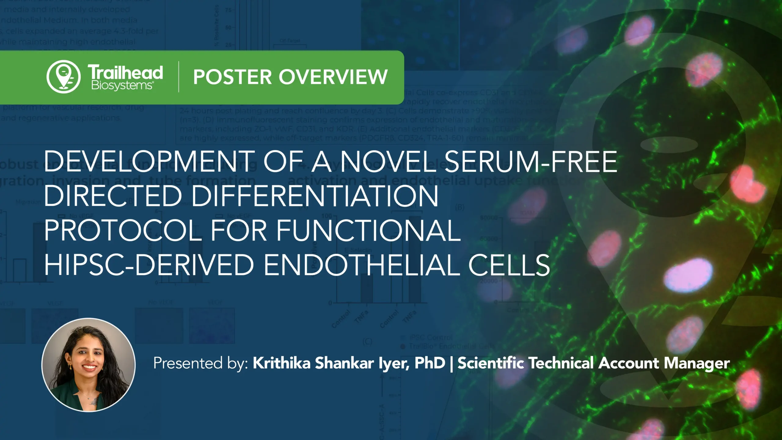
Development of a Novel Serum-free Directed Differentiation Protocol for Functional hiPSC-derived Endothelial Cells
Presenter:
Kirthika Shankar Iyer, PhD
Technical Account Manager, Trailhead Biosystems
Objective:
To develop a reproducible, serum-free iPSC differentiation process that generates highly pure, functional iPSC-derived endothelial cells suitable for scalable use across disease modeling, drug discovery, toxicity screening, regenerative medicine and tissue engineering.
Why Endothelial Cells?
Endothelial cells are essential regulators of vascular function, mediating transport, inflammation and tissue homeostasis across a wide range of biological systems. Leveraging iPSC-derived endothelial cells enables more predictive disease models and drug screening platforms, thereby improving the study of vascular biology and therapeutic development.
Video Transcript
Hi everyone, My name is Kritika Shankar Iyer from Trailhead Biosystems and today I will be presenting our work on the development of a novel serum-free directed differentiation protocol for generating functional iPSC derived endothelial cells.
Endothelial cells line the interior of blood vessels and play essential roles in regulating vascular function, inflammation, nutrient exchange, and tissue homeostatis.
Because of these functions, they are widely used in disease modeling, drug discovery, toxicity screening, regenerative medicine, and tissue engineering.
Our goal here was to develop a reproducible and scalable serum free differentiation process capable of generating highly pure functional endothelial cells that are ready for use immediately after thawing.
Starting with the differentiation process shown at the top of the poster, we use Trailheads’ high-dimensional design-of-experiments or HD-DoE platform to optimize a directed differentiation workflow.
This process guides iPSC through a mesoderm intermediate stage before generating endothelial cells.
Using a data-driven approach allowed us to improve reproducibility and reduce variability between production lots.
Next, we evaluated the identity and purity of the resulting endothelial cells.
The flow cytometric data shows that over 98% of the cells Co express CD31 and CD144, demonstrating a highly pure endothelial population immediately after thawing.
In the images to the right, you can see that the cells rapidly recover after thawing.
Within 24 hours they display characteristic endothelial morphology and by day three they form a confluent monolayer.
We also observed greater than 90% post Thor viability, indicating that the cells recover well from cryopreservation.
To further confirm endothelial identity, we performed immunofluorescent staining for markers including CD31, KDR, VWF, and 01.
The strong expression of these markers confirms both endothelial identity and maturation.
Additional flow cytometry analysis showed high expression of endothelial markers such as CD34, CD105, and KDR, while off-target markers associated with pericytes, epithelial cells, or undifferentiated stem cells remained minimal.
Together, these results demonstrate that the differentiation process produces a highly pure endothelial population that is ready-to-use immediately after thawing.
After confirming identity, we wanted to determine whether these cells function like endothelial cells.
In the migration assay shown in panel A self demonstrated approximately a 2.5 fold increase in migration in response to VEGF compared to unstimulated controls.
Migration is an important endothelial function because endothelial cells must move toward angiogenic signals during blood vessel formation and tissue repair.
Similarly, in the invasion assay, we observed approximately A4 fold increase in invasion and response to VEGF.
This indicates that the cells retain the ability to actively move through extracellular matrices, which is a critical step during angiogenesis.
We also evaluated tube formation on Matra gel.
As shown in panel C, the cells rapidly formed interconnected vascular-like networks with structures appearing within a few hours and becoming more extensive by 24 hours.
Taken together, these assays demonstrate robust endothelial functionality and angiogenic potential.
Next, we assessed whether the cells respond appropriately to inflammatory stimulation following treatment with TNF alpha.
We observed substantial increase in both E selectin and ICAM one expression compared to untreated controls.
This is important because endothelial activation during inflammation is characterized by the upregulation of these adhesion molecules, which facilitate immune cell recruitment to the sites of injury or disease.
We also observed increase ICAM one expression intensity, indicating that individual cells were expressing higher levels of this inflammatory marker.
In addition, over 98% of the cells demonstrated uptake of acetylated LDL AC LDL uptake is a hallmark endothelial function and reflects intact scavenger receptor activity and metabolic competence.
This provides additional evidence that these cells are not only expressing endothelial markers but are performing endothelial-specific functions.
Finally, we evaluated whether endothelial identity and function are maintained during expansion.
The cells expanded consistently over 3 passages while maintaining their characteristic cobblestone morphology.
Flow cytometry analysis showed that CD31 expression remained above 90% while PDGFR beta expression remained below 5%, indicating preservation of endothelial identity throughout expansion.
Importantly, expanded cells retained their ability to form vascular networks and continue to demonstrate ACLDL uptake, confirming that endothelial functionality is maintained during culture expansion.
In summary, we developed a serum-free HD-DoE-optimized differentiation protocol that generates highly pure functional iPSC-derived endothelial cells.
These cells demonstrate high viability after cryopreservation, robust endothelial functionality including migration, invasion, tube formation, inflammatory activation and Acldl uptake while maintaining their identity and performance through multiple passages.
Overall, TrailBio® Endothelial Cells provide a reproducible, scalable and physiologically relevant platform for vascular biology research, drug discovery, toxicity screening, and regenerative medicine applications.
Thank you.