Double Your Discovery: Buy One, Get One Free on Small Molecules

Webinar
EC Webinar | Presented on July 22, 2026
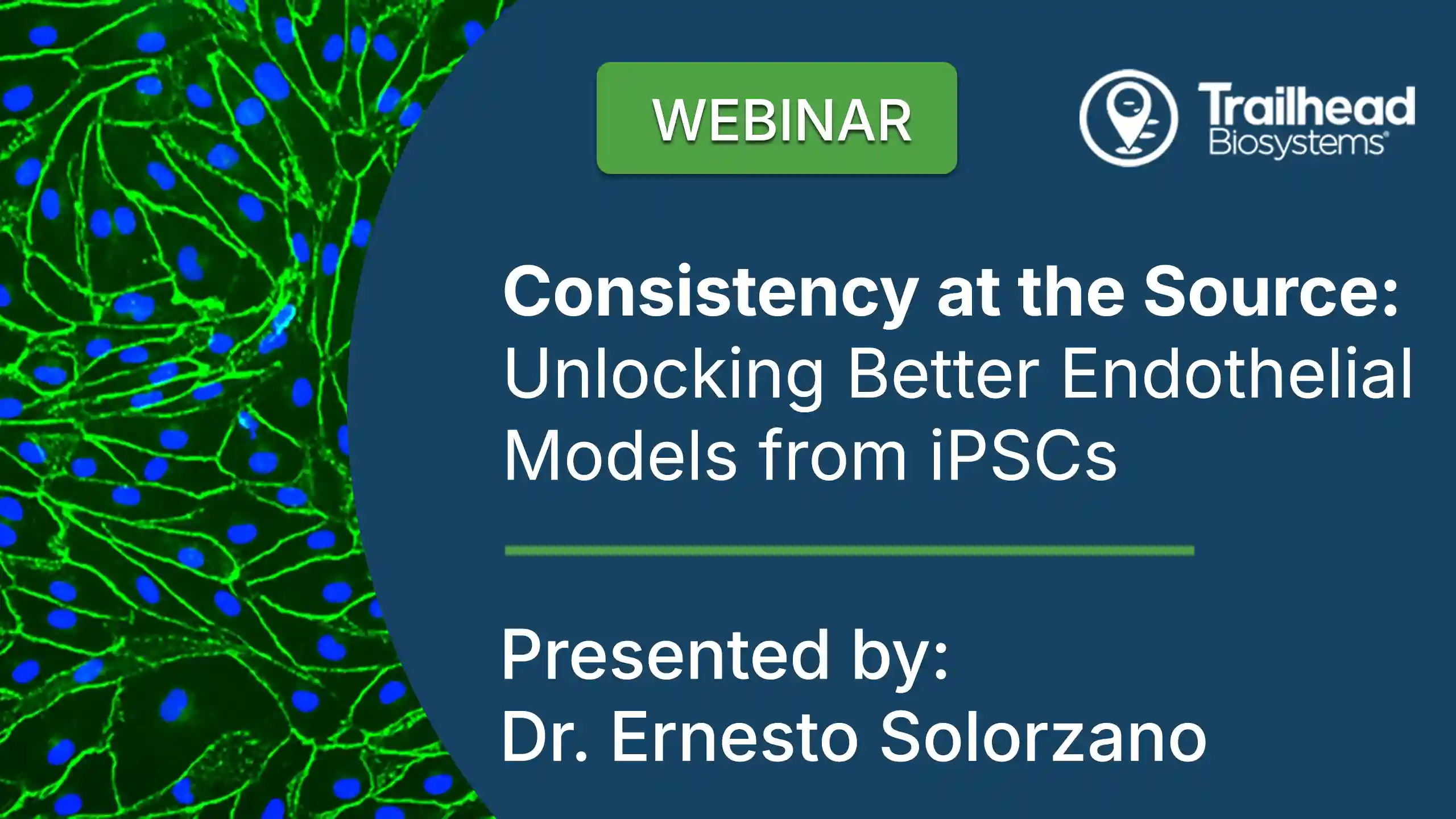
Consistency at the Source: Unlocking Better Endothelial Models from iPSCs
Presenter:
Ernesto Solorzano, PhD
Research Scientist, Trailhead Biosystems
Webinar Summary:
As the field advances toward New Approach Methodologies (NAMs), the need for consistent, human-relevant vascular models continues to grow. In this webinar, we’ll present a novel serum-free, directed differentiation approach for generating functional iPSC-derived endothelial cells.
We’ll discuss how controlled, reproducible cell production can reduce variability and enable more predictive models for drug discovery, toxicity testing, and vascular biology research.
Learning Objectives:
- Using HD-DoE® to map endothelial cell differentiation pathways
- Reducing variability compared to donor-derived primary cells
- Phenotypic characterization and functional validation of iPSC-derived endothelial cells
- Enabling more predictive vascular models across drug discovery and safety testing
Presenter Bio:
Ernesto Solorzano, PhD is a Research Scientist at Trailhead Biosystems and a member of the team behind the development of TrailBio® Endothelial Cells. His work focuses on creating robust, reproducible differentiation processes for human iPSC-derived cell products using Trailhead’s HD-DoE® platform. He earned his PhD from Northeast Ohio Medical University and has conducted research in vascular biology, stem cell differentiation, and therapeutic development.
Video Transcript
0:04
Hello everyone, and welcome to today’s webinar, Consistency at the Source, Unlocking better Endiothelial Models for IPSCS.
0:15
My name is Kevin Fluga, Lab Roots, and I’m going to be your moderator for today’s event.
0:19
Today’s Educational Web seminar is presented by Lab Roots and brought to you by Trailhead Biosystems.
0:26
We encourage you to participate today by submitting any questions you may have during the presentation.
0:31
To do so, simply type them into the Ask a Question box, which is located on the bottom of your screen, and click Submit.
0:39
We’ll answer as many of your questions as we have time for at the end of the presentation.
0:44
You can also use the Ask a Question box to let us know if you’re having any troubles seeing or hearing the presentation.
0:52
I’d now like to welcome our speaker for today’s webinar, Ernesto Solorzano, PhD, Research scientist at Trailhead Biosystems.
1:00
And a little bit about Ernesto.
1:02
He focuses on advancing human IPSC derived cell technologies for applications in disease modeling, drug discovery, and regenerative medicine.
1:12
He earned his PhD from Northeast Ohio Medical University, where he elucidated the osteopathic phenotype in complex lymphatic anomalies with a strong emphasis on translational science and moving innovation from bench to bedside.
1:28
Ernesto, we’re thrilled that you’re here with us today.
1:31
We’re greatly looking forward to your presentation.
1:33
So whenever you’re all set, I’m going to turn it over to you.
1:35
You can begin whenever you’re all ready.
1:40
Thank you for the kind introduction and I’m excited to share the work that we have done here at Trailhead.
1:46
1st, I’ll introduce Trailhead Biosystems and then touch on in Theo cell biology.
1:50
Next, we’ll look at why IPSE drive in Theo cells are valuable complementary options to donor sources.
1:58
I’ll then walk the trailheads, approach the IPSE drive in the Theo cells and expansion medium that we developed at Trailhead using our HDOE platform.
2:05
We’ll then follow up with presenting our newly developed expansion medium and finally over a pub with additional services offered at Trailhead.
2:15
Drill Head Biosystems was founded in 2015 and is based in Ohio and focuses on generating specialized cells derived from mesoderm, endoderm and actoderm.
2:25
Picture here is our facility which is located in Beachwood, OH just outside of Cleveland.
2:32
Since our mission is to create specialized cells from induced blue foot and stem cells, better known as IPS CS, it’s important to highlight where the real challenges in the field lie.
2:41
Many years of research have gone into reprogramming IPS CS, and we now know how to readily bring them back to a versatile state where they can become any cell type.
2:52
The real hurdle is in reprogramming.
2:54
It’s guiding these cells into the right specialized images.
2:58
That’s where childhood comes in.
3:00
Our workbook focuses on solving these differentiation challenges using IPS CS as a constable platform to generate functional specialized cells.
3:09
These cells can be then be applied across multiple areas from cell therapy, drug discovery, the disease modelling and tissue repair.
3:18
So let’s talk a little bit about some of the major challenges with IPSC derived cells today.
3:24
1st, the demand for high quality human cells is enormous, but right now the options are limited.
3:29
Researchers and companies alike are eager to use these cells for therapy, disease modelling, and drug discovery, but the field continues to run into the same problems.
3:38
Second, obtaining pure populations can be challenging.
3:41
Many of the available cells simply do not meet the standards needed for consistent and reliable results.
3:47
Also, there is a lack of consistency from batch to batch.
3:50
That means a researcher might not get one set of results from a bio and then something completely different from the next.
3:57
Fourth simply is a bottleneck.
3:59
These cells just aren’t available in the quantities that market truly demands.
4:03
And finally, much of the development and production is still being done manually.
4:07
This slows everything down and introduces opportunities.
4:10
Opportunities for error, which makes it very difficult to scale together.
4:14
These challenges create a major roadblock for the field, and solving them is the key to unlocking the full potential of IPC derives cells.
4:23
Now that we unlock a lot looked at the problems, let’s move into how we solve them.
4:28
At Trailhead, our approaches fundamentally change how stencil discovery and manufacturing are done.
4:33
Rather than relying on slow and traditional iterative processes, we leverage machine learning to enable the accelerated discovery.
4:40
This allows us to test and optimize conditions at a scale that simply isn’t possible by hand.
4:45
We also use this approach to efficiently explore the high dimensional space of regulatory inputs, the combinations of growth factors, small molecules, and cultural conditions that control sulfate.
4:56
By doing this, we can fine tune differentiation pathways in a way that drives both precision and consistency.
5:04
This result is vastly improved cell purity and the ability to generalize generate specialized subtypes that meet the needs of researchers would identify critical processes parameters.
5:15
These cells can then be manufactured reproducibly at a scale and because this entire system is built on flexible production platform, it can then be applied the multiple cell type or remaining cost effective.
5:27
The result is broader access, more reliable results and ultimately real impact for patients and the field as a whole.
5:36
So how do we actually make all of this happen?
5:39
The answer is the HRHDE platform.
5:42
HDDOE stands for High Dimensional Design of Experiments, and it’s the core technology that drives everything we do a trailhead.
5:48
Rather than rely on trial and error, we leverage proprietary in house software to design experiments with a level of skill and complexity that will be challenging to achieve through conventional approaches.
5:59
Our HD UE technology is built to lower the barrier of entry for researchers, whether through access to noble cell types, higher purities and streamline workflows, or lower research cost.
6:10
By pairing these designs with automated robotic execution, we can perform experiments with remarkable speed, precision, reproducibility, and scalability.
6:19
Much of the data generated through these experiments is novel, giving us a unique and expanding knowledge base.
6:25
And because everything is data-driven, we can determine critical processes parameters in an in an unbiased and empirical way.
6:33
This combination of advanced software’s, robotics and unbiased data is what allows us to exponentially accelerate the port cell protocol development and reliably generate specialized cell types.
6:46
So here is a visual example of our HDV platform.
6:49
We want to look at 12 factors on how they affect the cells.
6:53
Traditionally that will require about 4100 experiments.
6:58
With HDDOE, we can compress that large resource intensive 4100 experiments to 96, collecting the same amount of data, modeling that data and predicting the cell fate outcome.
7:08
This allows us to generate a lot of robust and reproducible protocols at an incredible pace.
7:15
So let’s move on to discussing the in the failure background which will be the topic for today And the Theo cells from the inner lining of vessels and serve a multi tool functions as summarized on this illustration.
7:28
While there are unique characteristics that can be attributed to the reciting organs and the Theo cells are often linked to the arterial vein or lymphatic subtypes, the primary functions of most of theocells are often linked to their barrier function, coagulation control, inflammatory response, and vascular tone regulation, and the Theo cells are are essential for studying the vascular system and have broad applications in vascular biology, disease modelling, drug discovery, and regenerative medicine.
8:00
Because many downstream assays depend on endothelial cell performance, high quality and well characterized cells are critical for generating reliable research results.
8:09
Here I will show primary endothelial cell data and how they are characterized in the literature.
8:14
In the upper left, we see two canonical endothelium markers, CD31 and von Willebrand factor or VWF.
8:21
CD31 plays an important role in endothelial barrier integrity, inflammatory signaling, and endogenesis, while VWS is involved in block clotting and serves as a marker of endothelium maturation.
8:35
Functional characterization is equally important.
8:38
In the lower left, ACLDL optic is used to assess the ability of endothelial cells to perform receptor mediated endocytosis AP, endothelial function.
8:48
Robust ACLDL uptake is a hallmark of functional endothelial cells.
8:54
In the center image 2 formation assays evaluate the cell’s ability to organize into capillary like networks in vitro, providing a measure of their androgenic potential.
9:04
Finally, the panels on the right demonstrate the endothelial inflammatory response following TNF alpha stimulation.
9:10
Endothelial cells increase expression of the addition molecules ICAM, which which promote leukocyte recruitment and addition.
9:21
Together, these markers and functional assays provide a comprehensive assessment of the endothelial identity, maturity, functionality, androgenic capacity, and responsiveness to inflammatory stimuli.
9:34
On this slide, I want to highlight the primary sources of endothelial cells and the limitations associated with each.
9:40
The most common donor sources are vessels from the umbilical cord and organ specific tissues.
9:45
These cells have been essential for research and clinical applications for decades, but both sources present challenges for umbilical cord derived cells.
9:53
The number of cells available from each donor is limited and there is significant variability in yield, quality and expansion potential.
10:03
While core blood core banks improve access, the overall supply remains finite.
10:08
Organ specific in the field cells present additional challenges during isolation, with protocols often varying by tissue type.
10:16
Further purification is typically required, and cell quality and proliferation capacity can differ substantially between donors.
10:23
Donor compatibility is another limitation.
10:25
Obtaining patient specific cells for autologous applications It’s not always practical, which restricts scalability.
10:33
In addition, creating large banks of genetically edited primary cells is challenging due to their limited lifespan and expansion capacity.
10:41
Together these limitations make it difficult to achieve the standardized scalability and reproducibility the modern research and industry require.
10:52
These challenges have driven interest in alternative cell sources, particularly IPSC derived endothelial cells, which offer the potential of an an essentially unlimited, standardized and engineerable supply of cells.
11:04
First, they bring consistency.
11:05
Because IPS CS can be banked and use it as a standardized material, differentiation runs can be can then be repeated under control protocols.
11:14
This minimizes lot to lot variability, which is a major issue when working with donor material where every lot can be behave different.
11:22
Second, I PS CS allow for customization.
11:25
You can generate lines for specific patients or engineer them to model genetic diseases, enabling disease specific applications.
11:32
Third, they provide an unlimited source of cells, unlike umbilical cord or organ tissues which are finite.
11:39
I PS CS can be expanded indefinitely in culture, ensuring A renewable and scalable supply of endothelial cells.
11:47
Another key benefit is reproducibility.
11:49
Standardized manufacturing produces consistent cell populations, making data more comparable across experiments, laboratories, and institutions.
11:58
IPS ES are also highly scalable.
12:01
They can be adapted to large scale production systems, making them valuable for research, high throughput drug screening, and preclinical testing.
12:08
Finally, their applications are broad.
12:10
IPS ES drives the TSS can be used the study vascular development, angiogenesis, vascular malformations, autoimmune disorders, inflammatory responses, and drug toxicity.
12:21
They also represent a promising platform for regenerative medicine and therapeutic development.
12:27
Importantly, IPSC interrogative cells align closely with the NIH’s increasing focus on the new approach methodologies better known as Naps.
12:35
As funding and regulatory agencies continue to prioritize human relevant, reproducible and translationary research models, IPSE based systems provide a scalable platform that can reduce reliance on donor tissue and improve the predicted value of preclinical studies.
12:50
Altogether, IPSE driven in cells provide a consistent, reproducible, scalable, enzymes aligned solution to many of the limitation limitations associated with donor derived cell sources.
13:05
While IPSC derivement Thea cells address many limitations of primary cells, their value ultimately depends on our ability to reliably generate functional endothelial cell populations in vitro.
13:16
During development and the Thea cells arise from the mesoderm through signaling pathways downstream of wind, VMP 4 and VEGF.
13:23
Most IPSC differentiation protocols may make these developmental cues guiding flurry button stem cells through a mesodermal stage and into endothelial lineages.
13:32
These products can also be tailored to generate arterial, venous and lymphatic in the TL cell subtypes.
13:36
For example, loop TF2 promote venous identity, whereas not signaling drives arterial specification.
13:43
There is still the resulting cells expressed canonical in the field markers such as CD31, KDR and VWF and exhibit key, exhibit key in the field functions including ACLDL uptake as shown in the image from Tennitol on this slide.
14:04
So let’s move on to the Trailhead approach for differentiating Trail bio endothelial cells.
14:11
Now that we’ve discussed the biology and background of endothelial cells, let’s look at how Trailhead generates endothelial cells from IPS ES.
14:18
Our process follows A2 stage workflow starting with the important stem cells with first driving towards Musaderm, then guide these cells into the endothelium.
14:31
Stage 2 represents our critical stage 4 in the field differentiation, where we develop high expression of CD31144 and KDR.
14:40
To optimize this transition, we apply our prior proprietary HCDOE platform.
14:46
This approach systematically evaluates combinations of small molecules and proteins to identify conditions that precisely control signaling pathways using differentiation.
14:57
By adjusting the timing and concentration of these signals, we can carefully guide these cells into the endothelial state.
15:04
In total, more than 70 factors were screened during the development of this process.
15:08
The result is a highly robust and reproducible differentiation protocol that reflects the steps of embryonic development while still providing the scalability and control needed for research and translational applications.
15:20
This ultimately enables reliable production of high quality endothelial cells at the end of stage 2.
15:28
At stage 2, mesoderma cells are are cultured in our optimised stage 2 medium, which promotes the expression of genes in supermarkets associated with the in the field of specification.
15:38
Following such specifications, we expanded ourselves prior prior to prior preservation.
15:44
Our protocol consistently generates highly pure in the field populations, which with greater than 95% of cells expressing both CD 31 and 144, also known as the CADIRI.
15:55
As demonstrated by flow cytometry, CD144 is a hallmark marker of mature endothelial cells and play a critical role in maintaining endothelial cell to cell junctions and vascular barrier integrity.
16:06
Importantly, our FROM formulations also minimize the presence of common contaminating cell types less than 5% of the population expressing the parasite marker CD140B or the epithelial marker CD324, indicating a highly enriched and the failure of culture.
16:22
In addition, the cells maintain approximately 95% viability immediately prior to prior preservation, demonstrating the high priority was achieved without compromising cell health.
16:34
Together, these results demonstrate the effectiveness of our HDDOE workflow and producing highly pure viable I PST derived in the Theo cell populations.
16:44
However, generating high quality cells is only part of the challenge.
16:49
Equally important is ensuring that this quality is maintained after prior preservation.
16:54
With that in mind, let’s take a look at the performance characteristics of Trail Bio and Theo cells and what you can expect when using them in your workflows.
17:05
Here we have a brief overview of the Trail buy on the Theo cell product.
17:10
Our product is released with several key specifications.
17:13
Each vial contains a minimum of 1,000,000 cells with greater than 80% hostile viability and greater than 90% expression of in the Theo marker CD31, CD 144 and KDR.
17:24
In addition, we maintain low levels of contaminated cell populations with minimal expression of parasite and epithelial cell markers.
17:32
These specifications ensure that the researchers receive a highly pure endothelial population with strong viability and a consistent phenotype right out of the vial.
17:41
Taken together, Trail Bio and filler cells are designed to be ready for use immediately after thaw, providing reliable and reproducible starting material for short term studies and long term applications.
17:55
Following thaw and three days of expansion, we extensively characterize the cells by flow cytometry to assess both phenotype and purity.
18:06
First, the cells demonstrated A robust proliferation over the 72 hour culture.
18:10
And exhibited the classical spindle morphology characterized of the healthy endothelial cells.
18:15
2nd trail by endothelial cells maintained strong expression of key endothelial markers with greater than 90% positivity of CD31, CD 144, CD three O 9, also known as a regibar or KDR, as well as CD3 to four and CD105.
18:34
Together, these markers confirm endothelial identity, they’re forming capacity, budget, responsiveness and androgenic potential.
18:42
Importantly, expression of contaminating cell markers remain low with minimal detection of parasite and epithelial populations.
18:50
We also observed very low expression of flare body marker trial 160, demonstrating efficient differentiation and no residual IPS CS Sticking together, these results show that trail bio endothelial cells not only retain their endothelial phenotype following prior preservation, but also rapidly recover fuller freight and reach confluence within just three days of culture.
19:13
We further characterize the endothelial identity of our cells using extensive immunocytochemistry analysis and ACLDL functional assays, whereby endothelial cells demonstrate robust expression of endothelial marker CD31, VKTRN, and KDR, confirming the flow cytometry results previously presented.
19:33
In addition, positive staining of ETS1 and UA1 provide further evidence of endothelial image commitment.
19:42
We also observe clear membrane localization of cell one, indicating the formation of material cell to cell junctions and embed reforming endothelial monolayer Expression of one wheel of RAM, where known as VWF, further supports the majority of the maturity and functional status of these cells beyond marker expression trail by on the cells the most demonstrate greater than 98% uptake of a CLDOA key functional characteristic of healthy cells and a widely accepted measure of endothelial activity.
20:14
Taking together, these data provide strong evidence that TRAIL bio endothelial cells exhibit both the molecular and functional characteristics expected of high quality endothelial cells, closely reassembling their primary endothelial cell counterparts.
20:30
Next, we sought to further evaluate the functional properties of our endothelial cells, especially their ability to migrate, invade, and form vascular networks.
20:38
All key features of angiogenic cells.
20:41
Angiogenic endothelial cells.
20:43
1st, we assess cell migration using a transfer assay in response to Vajab stimulation.
20:48
Joe by on the Theo cells demonstrated A robust chemotactic response with approximately a 2.5 Volt increase in migration compared to unsimulated controls as visualized by crystal Violet staining.
21:01
We then evaluated invasive capacity using a similar transfer system pre coded with H gel to mimic extracellular the the extracellular matrix barrier.
21:11
On their VED F stimulation, the cells exhibited strong invasive behavior resulting in approximately A4 fold increase in invasion relative to an all VED F condition.
21:21
Finally, we perform a tube formation essay, one of the most widely used functional assessments of endothetia cells.
21:27
When seeded into matrogen, Robi anesthetia cells rapidly organized into interconnected capillary like network, demonstrating their ability to recapitulate a key step of endogenesis in vitro.
21:40
Taking together, these results demonstrate that TRAIL bio endothelial cells are not only phenotypically endothelial cells, but also functionally active, exhibit the migration, invasion and vascular network formation capabilities expected of healthy androgenic endothelial cells.
21:58
We then evaluate the inflammatory response of our endothelial cells, a key functional characteristic of vascular endothelium whereby endothelial cells were stimulated with TNF alpha and analyzed for the expression of E selecting and Nikon one in the in the filial adhesion molecules that are up regulated during inflammatory and play inflammation and play critical roles in leukocyte recruitment and trafficking across the vascular wall.
22:23
Compared to untreated controls, TNF alpha stimulation induces a strong inflammatory response resulting in E selecting expression in approximately 43% of the population, while ICAM 1 is already expressed in our cells, similar to Cubex, we observed a strong increase in ICAM 1 levels that’s demonstrated by increased mean fluorescence intensity, also known as MFI, as shown through flow cytometry.
22:45
These findings demonstrate that TRAIL Bionithila cells remain responsive to inflammatory stimuli and are capable of recapitulating the activation state observed in native vascular endothelium.
22:59
Lastly, because endothelial cells are often expanded for downstream applications, we evaluated whether TRAIL by endothelial cells will maintain the phenotype and functionality over multiple passages.
23:11
As shown in the cumulative expansion graph, the cells were successfully expanded to three passages, achieving an average 4 fold expansion at each passage.
23:19
Importantly, endothelial purity remain consistent through our culture with City 31 expression maintained above 90% and CD140B expression remain below 10%.
23:31
Beyond marker expression, the cells continue to display their correct characteristic in the field of morphology following expansion by passage 3.
23:39
Trail by and the field of cells retain their ability to form capillary like networks into permission essays and maintain robust ACLDL uptake, demonstrating preservation of key endothelial functions.
23:52
Taken together, these results show that trail bio endothelial cells not only meet the hallmark criteria of functional endothelial cells, including canonical marker expression and eugenic activity and inflammatory responsiveness, but also maintain these defining properties through continuous passaging.
24:07
The stability makes them a reliable and versatile platform for vascular biology, disease modeling, and drug discovery.
24:14
Replications.
24:17
To conclude, TRAIL by Endothelial cells were developed to address the key challenges researchers face when working with endothelial models, purity, consistency, functionality and cost.
24:28
These cells were recently launched and are commercially available now.
24:31
First, our cells are highly pure with more than 90% of the population expressing key endothelial markers, including city 31 and City 144, while maintaining minimal levels of contaminating cell types.
24:45
2nd, they are truly ready to use.
24:48
Researchers can use these cells immediately following thaw or expand them in culture for up to three passages while maintaining their endothelial phenotype and functional performance.
24:57
Consistency is another major advantage.
25:00
By levering our HDA platform during development, we were able to optimize differentiation conditions, minimize variability, and generate a highly reproducible endothelial product beyond marker expression.
25:12
Trail Bio Endothelial cells have been extensively functionally validated.
25:17
We demonstrated robust ACLDL uptake, migration, invasion and to permission as well as inflammatory responses, confirming the key characteristics expected of healthy and fuel cells.
25:29
Finally, we affect the final the efficiency of our HDDOE workflow not only accelerated development but also reduce manufacturing costs.
25:38
This allows us to provide researchers with a high quality IPS to drive in fuel cell product at a more accessible price point.
25:46
I also want to take this opportunity to introduce you to our recently developed trail bio and the field expansion media.
25:55
Up to this point, all the data presented we’re generating using commercially available serum containing and the field medium.
26:02
However, as the field continues to move toward more reproducible and clinically relevant work flows, there’s an increased need for serum free culture systems.
26:11
Serum free media help reduce lot to lot variability, improve experimental consistency and support future translation into therapeutic and manufacturing applications.
26:20
To address this need, we developed the TRAIL Bile and the FILO Expansion medium, a fully serum free solution optimized for the culture and expansion of IPSE derived and the FILO cells.
26:33
The medium features a simple basal and supplement format that is ready to use after mixing, with no additional additives required.
26:40
Once prepared, the medium remains stable for at least two weeks during routine endothelial culture.
26:46
Importantly, this medium supports the entire endothelial workflow.
26:50
The same formulation can be used for postal recovery routines at expansion to formation essays ACLDL optic studies, eliminating the need for multiple specialized media.
27:03
Trio bio Endothelial expansion medium has been extensively validated for short and long term use of the trail bio on the T cell culture and expansion through at least three passages.
27:14
When compared with serum containing medium used in earlier studies, the Trail bio expansion medium delivered significantly higher cell yields at every passage, resulting in more than a tenfold increase in the total number of cells available by passage 3.
27:31
By passage 3, the cells continue to exhibit their characteristic endothelial morphology and retain their ability to form robust capillary like networks into formation.
27:40
Essays.
27:42
Importantly, this enhanced expansion did not come at the expense of cell quality.
27:46
Throughout passaging, the cells consistently maintain high expression of endothelial marker CD31 and low expression of CD140B, also known as PDTFR beta, demonstrating preservation of endothelial.
27:59
These findings were further supported by functional analysis, with greater than 95% ACLDL update maintaining maintained throughout expansion or from the preservation of endothelial activity.
28:11
Taken together, this result demonstrate that Trail Bio endothelial expansion not only supports robust endothelial cell growth but also preserves the phenotype, purity, and functionality of the cells during extended culture.
28:25
To benchmark on the serial expansion medium, we compared it against leading serum free and serum containing commercial formulation using Trail Bio, and the serial cells expanded over 3 passages by passage 3, Trail Bio medium achieved significantly higher cell yields than both competitors.
28:45
While the serum free competitor initially performed similarly, its expansion rate declined over time, resulting in substantially lower yields.
28:54
This was further confirmed by proliferation analysis, which showed significantly high proliferation activity in sales cultured with the TRAIL by a medium.
29:05
Morphological assessment at passage 3 supported these findings, with serum free competitor failing to reach the expected cell confluence, consistent with its reduced expansion of performance.
29:16
We also evaluated hemophilia identity and culture.
29:20
CD31 expression remain consistently high in cells cultured in the Trail by a medium, while the serum free competitor should significantly reduce CD 31 levels by passage 3IN contrast, the serum containing competitor exhibited progressively increased expression of the parasite marker CD140B across passages indicating enrichment of non endothelial populations.
29:46
Overall, these results demonstrate that the Trail Bio expansion medium delivers superior expansion, proliferation, maintenance of endothelial identity, and culture purity compared with commercially available serum free and serum containing alternatives.
30:03
After validating the Trail Bio expansion medium that’s the optimal condition for culturing or Trail bioendothelial cells, we sought to benchmark them against a commercial available IPSE, Dr.
30:13
and DSL competitor in their recommended medium.
30:18
Both cell products were cultured and analyzed in parallel through three passages.
30:23
Immediately following thaw TRAIL by on the field cells exhibit significantly higher CD31 expression, indicating a more enriched endothelial population at the starting point.
30:32
Following expansion, the competitor cells were able to reach comparable levels of CD21 expression, while CD1B levels remain generally similar between the groups across cell passages in terms of proliferation and cell yield.
30:48
Also, products demonstrated comparable performance, achieving robust expansion over the course of the study.
30:56
Additionally, by passage three, both populations maintain the characteristics of cobblestone morphology expected of healthy endothelial cells.
31:05
Overall, the most notable difference the the most notable differences between the two products was the higher endothelial purity observed Intel by endothelial cells immediately post op.
31:15
This provides researchers with a more consistent starting point population and reduces the need for additional recovery of enrich steps.
31:23
And finally, this these results achieved using our HDOE development platform, which enabled rapid optimization of the differentiation process while significantly reducing development and time cost.
31:35
This this efficiency allows us to offer a high quality in the Theta cell at a lower price point without compromising functionality, scalability and cell quality.
31:46
Finally, we evaluated the performance of our expansion medium in non IPSE derived cells to assess its broader applications across endothelial cell types.
31:55
We selected human umbilical vein in the Theta cells or Hubex as they are among the most widely used and well established in the field models in vascular research.
32:04
Following extended expansion, Ubex culture and Trail Bio medium maintains strong color for a capacity as an expected in the field morphology through Passage 3 and the field identity was also preserved with a robust expression of City 31, City 144 and KDR alongside low levels of parasite marker city 140B.
32:28
Finally, Hubecs remain highly competent at Passage 3, demonstrating efficient tube formation and greater than 99% ACLDL uptake.
32:38
Overall, these findings demonstrate that the Trail Bio expansion medium supports CUBEC growth while preserving the FILO phenotype and function during long term culture.
32:47
This gives us a standardized system to directly compare IPSE derived in FILO cells with Hubecs and we’re now extending these studies to additional endothelial cell lines to further evaluate the medium’s first ability.
33:01
To summarize, the TRAIL bio on the Silo expansion medium was assigned to support robust and reproducible endothelial cell expansion while maintaining cell quality and function.
33:11
Across our studies, the medium consistently generated higher cell yields than commercially available serum containing alternatives while providing the advantage of fully serum PRE formulations for a greater process control and consistency.
33:25
Importantly, expanded cells maintain key in the field characteristics and demonstrate A functionality across multiple assays supporting their sustainability for downstream applications.
33:36
We also validated the medium in Huex where it supports robust expansion, maintaining the field identity and pressure functional performance through extended passaging.
33:45
These results demonstrate the medium’s ability to provide a consistent and scalable culture platform across the verse and the field cell models.
33:53
Finally, the medium features Streamline 1 to 1 to 1 validation, Streamline 1 to 1, Basal 2 supplement format, simplifying workflow and inventory management.
34:06
Once prepared, the complete medium remains viable for at least three passages, providing at a convenience and consistency during routine culture.
34:14
The Trail Bio and the field expansion meeting will be available soon.
34:21
Let’s talk about other services offer that trailhead such as custom IPSE differentiation.
34:29
I want to briefly highlight our custom IPSE differentiation capabilities.
34:33
In addition to our off the shelf and the FILO products, Trabio also partners with researchers who want to generate cells from their own IPSE lines.
34:40
One of the major advantages of working with us is access to our executive platform, which allows us to rapidly identify and optimize the conditions needed to produce specific cell types in a scalable and reproducible way.
34:52
I know the key benefit is access to our internal expertise across all three germ layers, mesoderm and the derm and ectoderm.
35:00
Our team has extensively extensive experience designing differentiation strategies for a wide range of applications of specialized cell types.
35:10
This application allows researchers to work with patient derive or gene edited IPSC lines enabling disease relevant models while maintaining the consistency that comes from standardized differentiation workflows.
35:22
All together, this process, this provides a flexible way to generate customized cell populations while they’re leveraging thrill BIOS platform and expertise.
35:33
Here is a simplified overview of how a typical custom differentiation project works.
35:38
First, a project consultation where we discussed the research goals starting IPSC line and this RSL type next to the client provides line, provide line provided line undergoes sourcing and expansion to generate a stable population.
35:54
In the third step, our team performs optimization and differentiation, applying our HDOE platform and optimize protocol to get the cells into the desired lineage.
36:03
Finally, the cells undergo characterization and quality control before delivery with supporting data with the option for additional functional testing based on the project calls.
36:14
This workflow allows researchers to move from a starting IPSC line to a validated specialized cell population in a reproducible way.
36:24
Before we end this seminar, I want to briefly highlight a key few key takeaways.
36:29
First, our ICU platform gives us a powerful data-driven way to guide IPS CS into committed lineages with precision and reproducibility.
36:38
Second, our IPSC derived in the cells are highly pure, ready to use and then minimize love to love variation, making them a strong alternative to donor derived sources.
36:49
3rd, our data shows robust maintenance and an Athela identity with over 90% CD 31 CD 144 positive retain cells across 3 passages, low CD140B population and highly high hostile viability.
37:05
This finding support the use of our validated and Athela expansion medium, which will be available soon.
37:11
And finally, these cells can be easily adapted into more most work flows and applications.
37:16
This combination of innovation, quality and purity positions drill bio endothelial cells as a available tool for both research and translational work.
37:25
The cells are available for purchase now and the media kit will be launching soon.
37:34
With that, I conclude this seminar.
37:35
If you have any questions, please reach out to our sales team and support team.
37:40
We will now be moving on to the Q&A portion of the webinar.
37:43
I’m joined by my colleague Angelicas, our Scientific Director at Trailhead.
37:47
We’ll collect the development of these products and bring extensive expertise in vascular biology.
37:52
With that, we’re happy to take any questions.
37:57
Well, thank you very much, Ernesto for your informative presentation.
38:00
And as mentioned, we’re now going to start the Live Q and a portion of this webinar.
38:04
If you have a question you’d like to ask, please do so.
38:07
Now once again, just type your question into the Ask a Question box and click submit.
38:11
We’ll answer as many of your questions as we have time for and we already have a lot of great questions starting to filter in, so we’re going to jump right to it.
38:20
First one that was asked is what type of quality control assays are used to qualify these cells.
38:27
So hi Kevin, thank you for the introduction for a question harnessed or thank you for the presentation.
38:35
So yeah, what type of pilot controls are used to qualify ourselves?
38:38
So we conduct multiple assays to make sure that ourselves have high quality and ready to meet customer demands.
38:46
So from the essays that we do consistently, we do an analysis of viability, and we do that by flow cytometry.
38:55
We also measure the endothelial, endothelial purity of the cells by flow cytometry as well.
39:01
We’re not going to measure CD31 to make sure that they are of the endothelial identity and also CD140B to make sure that they don’t have contaminants.
39:10
And as a functional essay readout that we use, we use acetylated LDL to make sure that the cells are functional.
39:17
On top of that, you are guaranteed that you are going to receive more than 1,000,000 cells per vial.
39:23
So we conduct cell count analysis when we thought the cells and we guarantee that our cells are going to be negative for mycoplasma and also for microorganisms such as bacteria and full and fungus.
39:38
Our endometer we will have a particular QC, similar QC assessment to make sure that the media will make endothelial cells perform with the efficiency that you are looking for.
39:54
Excellent, thank you.
39:56
Next question, how is the batch to batch variation when comparing different batches?
40:03
OK.
40:03
The batch, batch of variation, one method that we use to assess the batch, batch variation was RNA SIC analysis.
40:10
So basically we send four different batches of sales for RNA SIC, 22 batches of RNT, 2 batches of manufacturing and we conduct A principal component analysis to see how close or how far together G cells cluster in our analysis and G cells cluster really close together.
40:33
They have indicating that they they have they are really similar.
40:37
There is not much batch to batch variability.
40:41
On top of the the RNAC analysis that we conducted, we also maintain the cells in culture in media for some time and we do assessments and on all these assessments the cells have consistence and they are reliable from the different batches.
41:03
So the batch, batch variabilities, it’s it’s similar, there is no batch, batch variability.
41:11
Awesome, thank you very much.
41:14
Have you tested these endothelial cells in Co culture systems with other vascular support cells?
41:22
There is a really good and interesting question but we haven’t done that yet.
41:28
Would be nice to combine these cells with fibroblasts and put them in a 3D culture essay to observe vascular formation, loomer formation, but we haven’t done that.
41:41
Additionally, another thing that we want to do is call culture these cells with other cell types such as liver cells or or neurons to make like organoids and see the the lumen and vascular vascularization of disorganoids in 3D culture systems.
42:04
OK, next question came in says we use large quantities of Hubix for screening studies.
42:11
Do you offer larger format lots or scaled up production beyond 1,000,000 cell vials?
42:20
Currently our standard offerings are 1,000,000 Salvio.
42:24
However, we acknowledge that some application require more cells, larger quantity of cells so we can work with customers on custom production requests and scale the manufacturing solutions with extended lead times.
42:40
OK, excellent.
42:42
Have you performed barrier function assays with the cells?
42:48
I haven’t performed that in house, but this is a interesting question.
42:55
And yeah, we can consider to do that in the future if the resources are available.
43:03
OK, excellent.
43:06
Moving right along here, how does your HDDOE platform make these endothelial cells different from other IPSC derived endothelial cells on the market?
43:17
So our platform accelerates process development by quickly evaluating large number of media components and identifying synergies, synergies, interactions between them.
43:28
So basically in one experiment we can evaluate multiple compounds at the same time, we can evaluate the interaction of these compounds and how these interactions influence the cell behavior.
43:40
So just allow us to optimize formulations more efficiently, remove unnecessary components and develop a robust differentiation protocols faster.
43:49
So as a result, we have a high quality and the Teta cell product with a more cost effective manufacturing process, helping us offer competitive pricing without compromising performance of the cells.
44:05
OK, excellent.
44:07
Well, thank you once again for your time today and your important research.
44:14
Want to thank Lab Roots and our sponsor, Trailhead Biosystems for underwriting today’s educational webcast.
44:20
Before we go, I’d like to thank the audience for joining us today and their interesting questions.
44:24
Questions that we did not have time for today and those submitted during the on demand.
44:29
Will be addressed by the speaker via the contact information you provided at the time of registration.
44:35
This webcast does have CME credits available upon completion of viewing the presentation.
44:41
After you watch the entirety of the presentation, please go back to the main micro site for this event and click the Paste CE button that is hyperlinked next to the CE credits.
44:52
From there, you’ll be prompted to fill out an evaluation form and from there you can obtain those credits.
44:58
This webcast can be viewed on demand.
45:00
Lab Roots will alert you via e-mail when it’s available for replay.
45:03
We encourage you to share that e-mail with your colleagues who may have missed today’s live event.
45:08
So once again, thank you both very much and until next time everyone, goodbye, bye.