The human brain is a complex and intricate organ, safeguarded by multiple layers of protection. Among these protective mechanisms is a family of specialized cells known as vascular leptomeningeal cells (VLMCs). VLMCS, also known as perivascular fibroblasts, are a family of fibroblast-like cells located within the protective membrane surrounding the brain and spinal cord. These cells were first identified in 2018 using single cell RNA sequencing (Vanlandewijck, Marques). The gene expression profile of VLMCs overlaps with that of mural cells (sharing expression of CD146/MCAM and CSPG4/NG2) and oligodendrocyte progenitors (sharing expression of PDGFRA).
This overlap in gene expression patterns with other cell types has made it difficult to distinguish VLMCs and study their functional role in the nervous system. Recently, ECM components such as DCN, LUM, COL1A1 and COL5A1 have been identified as VLMC signature markers. These markers suggest that VLMCs play a role in the establishment of ECM, cell adhesion and migration in the brain (Saunders, Ross, Rajan, Vanlandewijck). Moreover, two main populations of VLMCs have been identified, each with a unique gene expression profile. One subtype exhibits higher expression of ECM-related genes and angiogenesis markers, while the second subtype expresses membrane transport genes (Zeisel). This differentiation underscores the diverse functional roles VLMCs play within the brain’s complex environment.
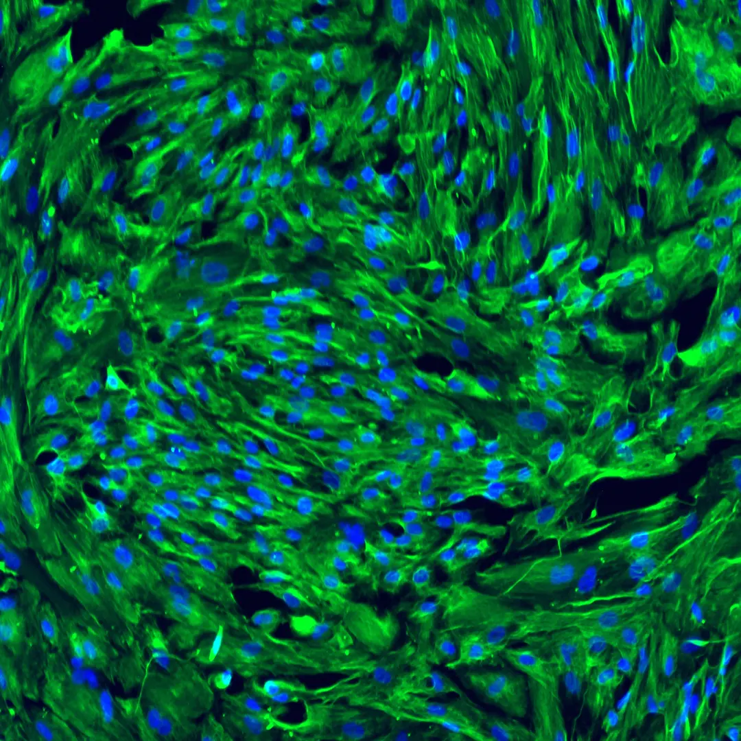
Morphology and Distribution of VLMCs Within the Blood-Brain Barrier
VLMCs not only express ECM components but also exhibit morphological differences compared to pericytes. High resolution images of the adult mouse brain have shown that pericytes possess longer processes than fibroblast-like cells (Rajan). Additionally, VLMCs have a distinct distribution in the brain. In vivo studies have identified these cells in the Virchow-Robin space (VRS) within the vasculature of the brain. The VRS is a fluid-filled area around the arterial and venous vessels that is involved in controlling penetration of molecules across the BBB into the brain (Dorrier). Unlike mural cells that are found around all vessel types, fibroblast-like cells have not been detected around capillaries (Bonney).
VLMCs loosely adhere to endothelial cells in the BBB while forming contact with end-feet of AQP4+ astrocytes. Other cell types in this region include pericytes and smooth muscle cells. Rajan’s studies demonstrated that fibroblast-like cells in this region contribute to vascular stabilization by depositing collagen proteins around the vessels. This role is integral, as these cells develop prior to the pericyte population (Ross). Historically mural cells are known to regulate blood flow and help stabilize nascent blood vessels by wrapping around the vessels (Ross). Moreover, it was shown that they can play a part in differentiation of pericyte progenitors (Rajan).
For more information about the BBB, see related post: What is the blood-brain barrier?
Putative Function of Vascular Leptomeningeal Cells in Blood-Brain Barrier Permeability
Brain fibroblast-like cells, including VLMCs, are thought to provide structural support by secreting ECM components, facilitating development of glymphatic system and recruiting immune cells in response to inflammation. Indirect evidence suggests that these cells can sense environmental changes such as blood flow variations in the perivascular space through mechanotransduction. They respond to these changes by gradually remodeling their environment and signaling to the neighboring cells, including endothelial and mural cells (Dorrier).
Fibroblast-like cells have interactions with other cell types in perivascular space (Sosa). One such interaction involves perivascular macrophages in waste clearance. It has been shown that loss of macrophages in the perivascular space leads to increased expression of ECM genes in fibroblast-like cells and a decrease in matrix metalloproteinase proteins. This results in the stiffening of vascular walls, which negatively affects waste clearance (Sosa, Drieu).
Vascular Leptomeningeal Cells’ Role in Brain Inflammation and Neuronal Diseases
In regions of inflammation, an immune cell niche is formed by recruitment of cells such as T cells and B cells to the affected area. These immune cells interact with fibroblast-like cells and are held together within the niche by ECM components secreted from fibroblast-like cells (Dorrier). This cellular network has been detected in areas of demyelination in multiple sclerosis.
Fibroblast-like cells also play a role in fibrotic scar formation in neuronal diseases. In conditions such as spinal cord injuries, traumatic brain injury and stroke, vascular leptomeningeal cells and their related ECM proteins influence the disease progression through regulation of signaling pathways such as IFN-gamma and SMAD, or through proliferation and higher expression of ECM proteins (Dorrier). Additionally, VLMCs, along with pericytes, become activated in response to stroke, undergo phenotypical and morphological changes and participate in regeneration of the blood-brain barrier at their progenitor state (Bernier).
The role of brain fibroblasts in regeneration of the BBB has also been demonstrated in brain injuries. The absence of fibroblasts in these cases resulted in enlarged injury volume and exacerbated BBB damage (Xu). In this context, fibroblasts promoted BBB repair through upregulation of tight junction proteins.
Conclusion
The scientific community is gaining an emerging appreciation for the role of vascular leptomeningeal cells, ranging from supporting the blood-brain barrier and facilitating cellular communication to responding to inflammation and injury. While the identification of specific ECM markers and distinct VLMC populations has provided valuable insights, the exact functions and roles of VLMCs are still being elucidated. Continued research is essential to fully understand the diverse roles these cells may play in brain physiology and neurological diseases.
References:
Bernier, LP., Hefendehl, J.K., Scott, R.W. et al. Brain pericytes and perivascular fibroblasts are stromal progenitors with dual functions in cerebrovascular regeneration after stroke. Nat Neurosci (2025). https://doi.org/10.1038/s41593-025-01872-y
Bonney SK, Sullivan LT, Cherry TJ, Daneman R, Shih AY. Distinct features of brain perivascular fibroblasts and mural cells revealed by in vivo two-photon imaging. J Cereb Blood Flow Metab. 2022 Jun;42(6):966-978. doi: 10.1177/0271678X211068528. Epub 2021 Dec 20. PMID: 34929105; PMCID: PMC9125487.
Dorrier CE, Jones HE, Pintarić L, Siegenthaler JA, Daneman R. Emerging roles for CNS fibroblasts in health, injury and disease. Nat Rev Neurosci. 2022 Jan;23(1):23-34. doi: 10.1038/s41583-021-00525-w. Epub 2021 Oct 20. PMID: 34671105; PMCID: PMC8527980.
Drieu A, Du S, Storck SE, Rustenhoven J, Papadopoulos Z, Dykstra T, Zhong F, Kim K, Blackburn S, Mamuladze T, Harari O, Karch CM, Bateman RJ, Perrin R, Farlow M, Chhatwal J; Dominantly Inherited Alzheimer Network; Hu S, Randolph GJ, Smirnov I, Kipnis J. Parenchymal border macrophages regulate the flow dynamics of the cerebrospinal fluid. Nature. 2022 Nov;611(7936):585-593. doi: 10.1038/s41586-022-05397-3. Epub 2022 Nov 9. PMID: 36352225; PMCID: PMC9899827.
Marques S, van Bruggen D, Vanichkina DP, Floriddia EM, Munguba H, Väremo L, Giacomello S, Falcão AM, Meijer M, Björklund ÅK, Hjerling-Leffler J, Taft RJ, Castelo-Branco G. Transcriptional Convergence of Oligodendrocyte Lineage Progenitors during Development. Dev Cell. 2018 Aug 20;46(4):504-517.e7. doi: 10.1016/j.devcel.2018.07.005. Epub 2018 Aug 2. PMID: 30078729; PMCID: PMC6104814.
Rajan AM, Ma RC, Kocha KM, Zhang DJ, Huang P. Dual function of perivascular fibroblasts in vascular stabilization in zebrafish. PLoS Genet. 2020 Oct 26;16(10):e1008800. doi: 10.1371/journal.pgen.1008800. PMID: 33104690; PMCID: PMC7644104.
Ross JM, Kim C, Allen D, Crouch EE, Narsinh K, Cooke DL, Abla AA, Nowakowski TJ, Winkler EA. The Expanding Cell Diversity of the Brain Vasculature. Front Physiol. 2020 Dec 3;11:600767. doi: 10.3389/fphys.2020.600767. PMID: 33343397; PMCID: PMC7744630.
Sosa MJ, Shih AY, Bonney SK. The elusive brain perivascular fibroblast: a potential role in vascular stability and homeostasis. Front Cardiovasc Med. 2023 Nov 24;10:1283434. doi: 10.3389/fcvm.2023.1283434. PMID: 38075961; PMCID: PMC10704358
Vanlandewijck M, He L, Mäe MA, Andrae J, Ando K, Del Gaudio F, Nahar K, Lebouvier T, Laviña B, Gouveia L, Sun Y, Raschperger E, Räsänen M, Zarb Y, Mochizuki N, Keller A, Lendahl U, Betsholtz C. A molecular atlas of cell types and zonation in the brain vasculature. Nature. 2018 Feb 22;554(7693):475-480. doi: 10.1038/nature25739. Epub 2018 Feb 14. Erratum in: Nature. 2018 Aug;560(7716):E3. doi: 10.1038/s41586-018-0232-x. PMID: 29443965.
Xu L, Nirwane A, Xu T, Kang M, Devasani K, Yao Y. Fibroblasts repair blood-brain barrier damage and hemorrhagic brain injury via TIMP2. Cell Rep. 2022 Nov 22;41(8):111709. doi: 10.1016/j.celrep.2022.111709. PMID: 36417884; PMCID: PMC9769996.
Zeisel A, Hochgerner H, Lönnerberg P, Johnsson A, Memic F, van der Zwan J, Häring M, Braun E, Borm LE, La Manno G, Codeluppi S, Furlan A, Lee K, Skene N, Harris KD, Hjerling-Leffler J, Arenas E, Ernfors P, Marklund U, Linnarsson S. Molecular Architecture of the Mouse Nervous System. Cell. 2018 Aug 9;174(4):999-1014.e22. doi: 10.1016/j.cell.2018.06.021. PMID: 30096314; PMCID: PMC6086934.